Organization of treatment and rehabilitation in Germany.
Complete medical support.
Complete medical support.





+49 176 17917001
All departments
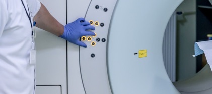
Positron emission tomography (PET) is a nuclear medicine imaging method that produces a series of tomographic images of the entire body or a part of it. The resulting images visualize the area under investigation layer by layer.
PET scanning uses radioactive pharmaceuticals, most commonly fluorodeoxyglucose, which are distributed throughout the body when administered, accumulating in those parts that have a particularly active metabolism. Tumors and metastases have precisely this elevated metabolism, they consume more glucose than healthy cells. The measuring unit in the PET device registers the radiation from the radioactive pharmaceutical in these areas, and the connected computer uses this data to process images.
Unlike other imaging methods such as radiography, CT or MRI, which visualize bones, tissues and organs, PET shows the metabolic activity of tissue, since the administered radioactive substances become part of the body's metabolism.
PET scanning resembles scintigraphy, in which radioactive substances are also introduced into the body.
PET scanning is very accurate. This type of diagnostics allows detecting changes in tumor tissue at early stages, even if their size is only 1 mm, and visualizes not just malignant neoplasms, but also metastases, showing their exact location.
 +49 176 17917001
Or send us a message: